Since the discovery of X-rays, medical imaging technology has begun to change the way doctors diagnose and treat patients. Due to continuous technological advances, modern medical imaging devices are capable of performing highly accurate diagnoses. The two most important aspects of modern medicine are diagnosis and treatment. Diagnosis is the premise of medical treatment, and treatment is fundamentally based on diagnostic results. The accuracy of the diagnosis result will influence the success of the treatment. We need to learn about these medical imaging devices and understand how they differ from the technology they use.
What are Medical Imaging Devices?
Medical imaging devices are widely used in clinical diagnostic equipment. They are devices that reproduce the structure of the human body as images by using different media. The image information corresponds to the actual structure of the human body in terms of spatial and temporal distribution.

According to the imaging principle, medical imaging devices can be divided into X-ray imaging devices, magnetic resonance imaging (MRI) devices, computed tomography (CT scans) devices, nuclear medicine imaging devices including positron-emission tomography (PET), ultrasound imaging devices, optical imaging devices, thermal imaging devices, etc. The imaging principles of various medical imaging devices are different, and their clinical application characteristics are not exactly the same. Different devices can only complement each other rather than replace each other. No one type of imaging device is always the best. Each of them has its advantages and disadvantages. Below we will discuss the features, advantages and disadvantages of several imaging devices.
What Types of Technology Are Used in Medical Imaging Devices?
As mentioned above, there are different technologies used in medical imaging devices. Each type of imaging technology will deliver different image information of the body. They can be used to check the possible injury, disease, or effectiveness of the medical treatment. Firstly, let’s learn the working principles of these technologies.
1. X-ray Imaging
Among various types of high-precision medical devices in medical institutions, medical diagnostic X-ray equipment is a medical imaging inspection means that has been applied earlier and has wide clinical popularity. With its large amounts of information and strong spatial analysis ability, X-ray imaging has obvious advantages in examining dynamic and subtle lesions.
Medical diagnostic X-ray devices are mainly composed of an X-ray generator, an X-ray image receptor, an image processor, and X-ray accessory and auxiliary devices. Its imaging principle is based on the use of X-rays that enter the body and produce an ionizing effect that causes a change in biological properties. This is then transformed into a visual image through a series of actions.
Medical diagnostic X-ray devices have the irreplaceable role of other imaging equipment in the clinical diagnosis of certain diseases. With the development of technology and the emergence of other imaging technologies, clinical requirements for medical diagnostic X-ray devices are higher. For example, safe radiation dose and protection, diversified high-definition image quality, digital image processing system, etc. The development of medical X-ray diagnostic technology has always been aimed at reducing irradiation dose and improving image quality. The integration of medical diagnostic X-ray equipment with other imaging technologies and the development in the direction of digitization, high frequency, and intelligence is an inevitable requirement for clinical development.
2. Computed tomography (CT Scans)
CT is one of the most common medical imaging devices in clinical applications, which has an important role in medical diagnosis. Its scanning part consists of an X-ray tube, detector, and scanning frame. During the scanning process, the X-ray emitter and receiver in the scanning frame rotate at high speed around the axis of the scanning frame, while the scanning frame drives the object to be scanned through the X-ray plane and scans the object. The information collected after scanning will be stored and calculated by a computer system. Finally, the computer-processed, reconstructed image will be displayed on the screen through the image display and storage system.
With its features of fast scanning time and clear images, CT can be used for the examination of a wide range of diseases and has been widely deployed in hospitals around the world. The principle of CT scans is to scan a certain thickness of the human body part with an X-ray beam, which is then received by a detector, transformed into an electrical signal by a photoelectric converter, and then converted into a digital signal by an analog-digital converter to a digital signal.
3. Magnetic Resonance Imaging (MRI)
Magnetic Resonance Imaging is a technology that uses the magnetic resonance signal of nuclei (mainly hydrogen protons) in water molecules in the human body for tissue or organ imaging via reconstruction in a strong magnetic field. The magnetic resonance imaging system mainly consists of the main magnet system, gradient magnetic field system, radio frequency system, computer processing system, and auxiliary equipment. Magnetic resonance imaging has the advantage of high resolution for soft tissues and no radiation damage. It is easy to detect lesions and show features and can be used in multiple sites, especially in the central nervous system.
4. Ultrasound
Ultrasound imaging is an imaging technique that uses the physical properties of ultrasound and the acoustic parameters of human tissue. The vibration frequency of the sound source used for ultrasound diagnosis is generally 1-10 megahertz. Compared with other imaging technologies, ultrasound imaging is safe and non-invasive, painless for patients, has good real-time performance, and low price. It has a high value in the prevention, diagnosis, and treatment of diseases. Ultrasound imaging is widely used in gastroenterology, gynecology, obstetrics, urology, thoracic, small organs, pediatrics, cardiology, emergency medicine, and other clinical examinations, and gradually combined with other clinical departments.
5. Position Emission Tomography (PET) Imaging
Positron emission tomography, which belongs to nuclear medicine imaging, is the most important new technology that has emerged in the field of nuclear medicine in the last 20 years. PET scans are widely used in the areas of oncology, cardiovascular disease, and neuroscience to detect and evaluate diseases such as heart disease and epilepsy.
PET scans use a small dose of a chemical: a radionuclide combined with glycosylation. The compound is injected into the patient, and the radionuclide emits positrons. The PET scanner then rotates around the patient’s head to detect the positron emitted by the radionuclide. Since malignant tumors grow extremely fast compared to healthy tissue, tumor cells consume more carbohydrates, to which radionuclides are attached. So the computer uses the measurement of the used carbohydrate to produce a color-coded image. If radionuclides build up in certain areas, it could be a sign of disease.
A Comparison of Different Medical Imaging Technologies
Below are the different types of imaging and an explanation of how they are used, as well as some of the advantages and disadvantages of each.
| Imaging Type | Advantages & Disadvantages |
| X-ray Imaging X-rays are suitable for areas or organs with good natural density contrast, mainly for displaying images of bones, and some dense objects. They are often used to diagnose fractures. But it can also be used to detect the development of diseases such as pneumonia and cancer. | Advantages · Most common & widely available · Relatively cheap and fast imaging · Painless and non-invasive Disadvantages · Exposure to X-ray (ionizing radiation), causes a very small risk of cancer in future |
| Computed tomography (CT scans) CT scans generate 3D images from multiple X-ray beams, and doctors and patients can view internal body structures such as lung disease, bones, tumors, etc. | Advantages · Non-invasive, painless, and fast imaging · Offer more information than X-ray imaging · Detect or exclude whether there are more serious problems Disadvantages · Higher radiation risk than standard X-rays · Some people will have allergic reactions to contrast medium · Contrast medium may cause kidney problems |
| Magnetic resonance imaging (MRI) MRI uses magnetic fields and radio frequency waves to scan the body, and then create detailed images showing organs, soft tissues, and other areas through computers. | Advantages · Painless and non-invasive · No ionizing radiation · MRI provides a higher level of detail of internal soft tissue structures than CT scans Disadvantages · The process is longer and noisier · Requires the patient to remain stationary during the scan · Not suitable for people with claustrophobia, certain sedation measures are required before the examination · High cost |
| Ultrasound Ultrasound technology uses high-frequency sound waves to produce images. It can be used to examine organs, soft tissues, or fetal development. | Advantages · No ionizing radiation · Safe for pregnant · Usually non-invasive and painless Disadvantages · Sound waves do not pass well through bone or air · The quality of the image is easily influenced by operating techniques and other factors |
| Nuclear medicine imaging including positron-emission tomography (PET) The scanner displays images of different parts of the body by using gamma rays emitted by a tracer that is injected, inhaled, or taken into the body. It is commonly used to diagnose cancer, heart disease, and brain disorders. | Advantages Painless PET scans may help detect diseases that cannot be found on an MRI or CT scan Disadvantages · Exposure to gamma raysPain or redness when injecting a tracer · Not suitable for people with claustrophobia, if a PET examination is to be done, certain sedation measures are required |
Current status of Medical Imaging Devices
The medical imaging device is a major market in the medical device industry. The global medical imaging market will be approximately $28 billion in 2021 and is expected to approach $47.4 billion in 2030, growing at an estimated CAGR of 4.9% from 2022 to 2030. (Date via Grand view research)
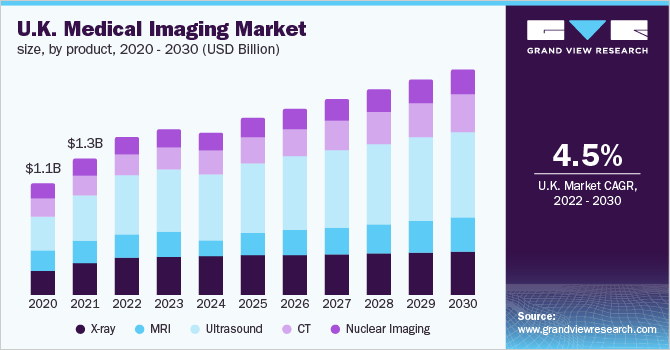
Medical imaging device is also the segment with the highest technological barriers in the medical device industry. Medical imaging is a typical multidisciplinary industry. The global market has long been an oligopoly situation. GE Healthcare, Philips, Siemens, and some Japanese manufacturers have accumulated deep patents and technologies. And the global medical imaging core component production technology is also concentrated in the hands of a few enterprises.
